Integration of 4D-MRI into robust treatment planning for adaptive hadron therapy of pancreatic carcinoma
Project leaders:
Aswin Hoffmann, MSc PhD (Helmholtz‐Zentrum Dresden-Rossendorf, Institute of Radiooncology - OncoRay, Dresden, University Hospital Carl Gustav Carus at the Technische Universität Dresden, Dept. of Radiation Oncology, Dresden), aswin.hoffmann[at]oncoray.de
Dr. rer. nat. Asja Pfaffenberger (German Cancer Research Center (DKFZ), Medical Physics in Radiation Oncology (E040)), a.pfaffenberger[at]dkfz.de
Funded since: April 2016
With 16 000 annual fatalities in Germany, pancreatic carcinoma is the fourth deadliest form of cancer. Standard treatment includes surgery, chemotherapy and radiotherapy or – depending on the tumor stage – a combination of the above. The intrafractional movement caused by respiration of the patient as well as the intrafractional deformation of the tumor tissue in patients with pancreatic carcinoma pose a special challenge to radiation therapy. Physicians currently include fairly big safety margins in radiation planning to take the mobility of the tumor into account. This inevitably leads to high toxicity in healthy surrounding tissue. The patient-specific evaluation of tumor mobility and the planning of individualized safety margins could improve the effect of radiotherapy for patients with pancreatic carcinoma.
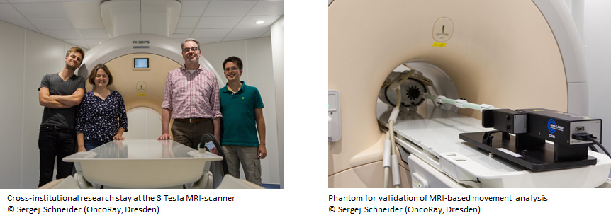
In this project a cross-institutional protocol for the examination via magnetic resonance imaging (MRI), which is optimized for diagnosis of pancreatic cancer, will be developed and used as the optimal basis for characterization of the relevant tumor volume and its deformation. Tumor mobility shall be extracted from time-resolved volumetric MRI (4D-MRI). The reduction and reproducibility of movements will be analyzed with different methods of immobilization (abdominal corset, vacuum mattress). Our hypothesis states that reduced tumor mobility during radiation and imaging can be achieved through an abdominal corset and quantified via MRI. The movement analysis via 4D computed tomography shows deficits and inaccuracy because of the low soft tissue contrast und sorting-artefacts. A 4D-MRI could overcome these issues. Finally the information about tumor mobility from 4D-MRI data shall be incorporated into radiation treatment planning of particle therapy for patients with pancreatic carcinoma.


